About
Microscopic Spine Surgery in Ahmedabad

Microscopic spine surgery in Ahmadabad is most recent, advanced and safest and involves surgery under guidance of microscope with the help of special tubular cylindrical retractor instruments which enhance the safety due to the better magnified 3 dimensional visualization obtained due to microscopic assistance. Spine and orthopaedic centre hospital which gives advantages of microscopic spine surgery in Ahmadabad, Gujarat, Memnagar, Gurukul, Sarkhej, Thaltej, S G highway, Vastrapur, Prahladnagar, Vasna, Bodakdev, Vejalpur, Ashram Road, Jodhpur, Ellisbridge, Mithkhali, Paldi, Chandkheda, Navrangpura, Sabarmati, C G Road, Wadaj, Ambawadi, Naranpura, Satellite, Usmanpura, Gandhinagar.
- Smaller incisions
- Less blood loss
- Less pain post operative
- Shorter hospitalisation
- Early recovery
- Less infection rates
- No long term morbidity
Treatment Option
With the advent of modern imaging technology, neuro navigation and microsurgical instrumentation several spine surgery that required large incision and open surgeries can now be done as minimally invasive procedures. These microscopic spine surgeries requires extremely small sized incisions cause minimal internal tissue damage require minimal hospital stay and enable a faster return to normal life there are some procedure that can be done using microscope surgery technique are listed below
- Minimally invasive spine fusion
- Microdiscetomy
- Microlaminectomy
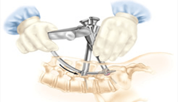
Spine surgery is performed
- A small incision in the midline of the low back A first the back muscles are lifted off the lamina arch of the spine since these back muscles run vertically they can move out of the way rather than cut.
- The surgeon is then able to enter the spine by removing a membrane over the nerve roots and uses either operating glasses or an operating microscope to visualize the nerve root.
- Often small portion of the inside facet joint is removed both to the facility access to the nerve root and to relive pressure over the nerve.
- The nerve root is then gently moved the side and the disc materials is removed from the under the nerve root


